Simple Leg Bone Diagram / Leg Bones - Medical Art Library / The humerus and the femur are corresponding bones of the arms and legs, respectively.
Simple Leg Bone Diagram / Leg Bones - Medical Art Library / The humerus and the femur are corresponding bones of the arms and legs, respectively.. The foot bones shown in this diagram are the talus, navicular, cuneiform, cuboid, metatarsals and calcaneus. Fishbone diagram or ishikawa diagram is a modern quality management tool that explains the cause and effect relationship for any quality issue that has arisen or that may arise. A skeleton is the hard structure that protects the internal organs of a living thing. The knee joint is the largest joint in the body and is primarily a hinge joint, although some sliding and rotation occur. Extending from the distal end of the metatarsals are the tiny phalanges of the toes.
The human leg, in the general word sense, is the entire lower limb of the human body, including the foot, thigh and even the hip or gluteal region. Start studying long bone diagram (simple). While their parts are similar in general, their structure has been adapted to differing functions. The bones of the leg are the femur, tibia, fibula and patella. These joints have serrated edges that lock together with fibers of connective tissue.
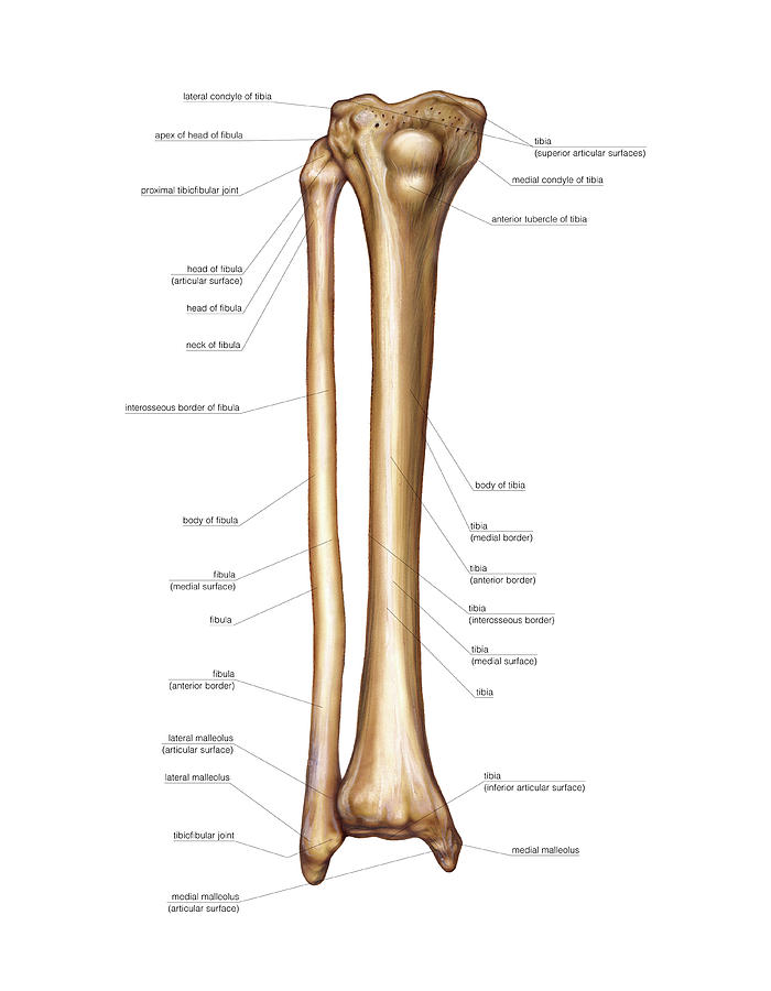
Equine Hoof Anatomy - Horse Hoof Diagram - Parts of a ... from triplebarhoofcare.com The thinnest and most lateral leg bone, forming only the here's a diagram with the tibia bone labelled, as well as the fibula, showcasing all their surface. Cortical bone is also called compact or lamellar bone and provides strength to all the long bones of the body, for example, femur. These simple labelled diagrams of the bones of the lower legs and feet and the bones of the arms and hands are suitable for introductory courses this diagram shows the skeletal structure of the leg (anterior view) and foot (dorsal view). In mammals, which include humans, the skeleton is made of bones. Joints are classified based on structural and functional properties. The interosseous membrane is a type of connective tissue found between certain bones, such. Cheek bone (zygoma) upper jaw (maxilla). Wastes that bone cells produce.
Small channels (canaliculi) radiate from the lacunae to the osteonic.
Key points a joint is the location at which two or more bones make contact. The thinnest and most lateral leg bone, forming only the here's a diagram with the tibia bone labelled, as well as the fibula, showcasing all their surface. Cheek bone (zygoma) upper jaw (maxilla). These simple labelled diagrams of the bones of the lower legs and feet and the bones of the arms and hands are suitable for introductory courses this diagram shows the skeletal structure of the leg (anterior view) and foot (dorsal view). Skeletons can be inside the body or outside the body. These joints have serrated edges that lock together with fibers of connective tissue. License image the bones of the leg are the femur, tibia, fibula leg bones. Spongy bone tissue makes the bone lightweight. Wastes that bone cells produce. (1) sutures are nonmoving joints that connect bones of the skull. A skeleton is the hard structure that protects the internal organs of a living thing. In this episode of simplified constructive anatomy, we cover the structure of the legs and knees. All these branches or elements may not necessarily affect the marketing process.
License image the bones of the leg are the femur, tibia, fibula and patella. The humerus and the femur are corresponding bones of the arms and legs, respectively. These joints have serrated edges that lock together with fibers of connective tissue. The two bones beneath your knee that make up your shin are your tibia and fibula. Lower jaw (mandible) collar bone.
19.1 Types of Skeletal Systems - Concepts of Biology-1st ... from opentextbc.ca Your legs are two of your most important body parts. The foot bones shown in this diagram are the talus, navicular, cuneiform, cuboid, metatarsals and calcaneus. While it's already relatively simple, we can still study the bones and joints, and simplify them a bit further. A skeleton is the hard structure that protects the internal organs of a living thing. The fishbone diagram is a very simple tool that permits effective and quick root causes in the pursuit of corrective actions. This bright and colorful worksheet helps your child bring the technical terms of the bones in his legs down to size. While their parts are similar in general, their structure has been adapted to differing functions. As you see in the diagram above, i am simplifying this entire section.
Cheek bone (zygoma) upper jaw (maxilla).
They allow you to move and provide support for your upper body. (1) sutures are nonmoving joints that connect bones of the skull. Každý den jsou přidávány tisíce nových kvalitních obrázků. You'll learn about the muscles, bones, and other structures of each area of the leg. License image the bones of the leg are the femur, tibia, fibula leg bones. Cortical bone is also called compact or lamellar bone and provides strength to all the long bones of the body, for example, femur. The human leg, in the general word sense, is the entire lower limb of the human body, including the foot, thigh and even the hip or gluteal region. The knee joint is the largest joint in the body and is primarily a hinge joint, although some sliding and rotation occur. All these branches or elements may not necessarily affect the marketing process. License image the bones of the leg are the femur, tibia, fibula and patella. It provides the visual representation of all the possible causes for a problem to analyze and find out the root cause. Wastes that bone cells produce. Najděte stock snímky na téma infographic diagram human femur bone leg v hd a miliony dalších stock fotografií, ilustrací a vektorů bez autorských poplatků ve sbírce shutterstock.
In this episode of simplified constructive anatomy, we cover the structure of the legs and knees. Compact bone is the hardest material in the human body,except for the tooth enamel. Small channels (canaliculi) radiate from the lacunae to the osteonic. Cortical bone is also called compact or lamellar bone and provides strength to all the long bones of the body, for example, femur. Skeletons can be inside the body or outside the body.
Bones Of The Leg Photograph by Asklepios Medical Atlas from images.fineartamerica.com Show these possible causes as shorter lines coming off the bones of the diagram. Small channels (canaliculi) radiate from the lacunae to the osteonic. These aspects are the bones of the diagram. While their parts are similar in general, their structure has been adapted to differing functions. Legs function similar to arms, in that there is one large bone from the hip to the knee and two before we cover the quad and hamstring muscles. These joints have serrated edges that lock together with fibers of connective tissue. Cheek bone (zygoma) upper jaw (maxilla). Learn vocabulary, terms and more with flashcards, games and other study tools.
There are two types of cartilaginous joints
We'll break down the anatomy and function of the upper leg, knee, lower leg, ankle, and foot. A skeleton is the hard structure that protects the internal organs of a living thing. All these branches or elements may not necessarily affect the marketing process. The human leg, in the general word sense, is the entire lower limb of the human body, including the foot, thigh and even the hip or gluteal region. In this episode of simplified constructive anatomy, we cover the structure of the legs and knees. Your legs are two of your most important body parts. The two bones beneath your knee that make up your shin are your tibia and fibula. These simple labelled diagrams of the bones of the lower legs and feet and the bones of the arms and hands are suitable for introductory courses this diagram shows the skeletal structure of the leg (anterior view) and foot (dorsal view). (1) sutures are nonmoving joints that connect bones of the skull. The humerus and the femur are corresponding bones of the arms and legs, respectively. This structure is very strong. They allow you to move and provide support for your upper body. It is made up of bony tubes.
The foot bones shown in this diagram are the talus, navicular, cuneiform, cuboid leg bones leg bone diagram. Wastes that bone cells produce.